Abstract
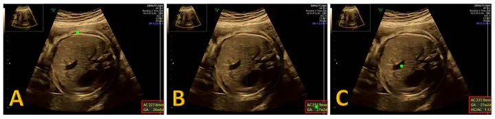
Objectives: Operators performing fetal growth scans are usually aware of the actual gestational age. This may lead to an expected value bias when performing biometric measurements. Methods: We prospectively collected full‐length video recordings of routine ultrasound growth scans coupled with operator eye tracking. The expected value was defined as the actual gestational age at the time of the scan. Expected value bias was defined as occurring when the operator looked at the ‘‘measurement box’’ during the process of caliper adjustment before saving a measurement. We studied the three standard biometric planes on which measurements are taken: Head Circumference (HC), Abdominal Circumference (AC), and Femur Length (FL). We evaluated the occurrence of expected value bias and quantified the impact of biased measurements. Results: We analyzed 272 third trimester growth scans with a total of 1409 measurements (354 HC, 703 AC, and 352 FL) performed by 16 operators. Expected value bias occurred in 91.4% of the saved standard biometric plane measurements (85.0% for HC, 92.9% for AC, and 94.9% for FL). The operator adjusted the measurement toward / away from the expected value in 48% /20% of the biased standard plane measurements (p<0.001). On average, measurements were corrected by 2.3 ± 5.6, 2.4 ± 10.4, and 3.2 ± 10.4 days of gestation towards the actual gestational age for the HC, AC, and FL, respectively. Additionally, we note a statistically significant reduction in measurement variance once the operator was biased (p=0.026). Comparing the lowest and highest possible estimated fetal weight (EFW), we note that the discordance, in percentage terms, was 10.1% ± 6.5% and that in 17% (95% CI 12‐21%) of the scans, the fetus could be considered as small‐for‐gestational‐age or appropriate‐for‐gestational‐age if using the smallest or largest possible measurements, respectively. Similarly, in 13% (95%CI 9‐16%) the fetus could be considered as large‐for‐gestational‐age or appropriate‐for‐gestational‐age if using the largest or smallest possible measurements, respectively. Conclusions: During routine third‐trimester growth scans, expected value bias frequently occurs and significantly changes measurements of standard biometric planes.
*L. Drukker and R. Droste contributed equally to this work.
BibTex
@article{doi:10.1002/uog.21929,
  author = {Drukker, Lior and Droste, Richard and Chatelain, Pierre and Noble, J. Alison and Papageorghiou, Aris T.},
  title = {Routine third-trimester growth scans: how common is expected value bias?},
  journal = {Ultrasound in Obstetrics \& Gynecology},
  volume = {in press},
  doi = {10.1002/uog.21929},
  url = {https://obgyn.onlinelibrary.wiley.com/doi/abs/10.1002/uog.21929},
  eprint = {https://obgyn.onlinelibrary.wiley.com/doi/pdf/10.1002/uog.21929}
}